Beschreibung
Produktdetails
Einband
Gebundene Ausgabe
Erscheinungsdatum
13.07.2026
Abbildungen
XIX, 209 p. 322 illus., 238 illus. in color.
Herausgeber
Dayong Gu + weitereVerlag
Springer SingaporeSeitenzahl
209
Maße (L/B/H)
24,1/16/1,8 cm
Gewicht
558 g
Auflage
1. Auflage
Sprache
Englisch
ISBN
978-981-9586-67-7
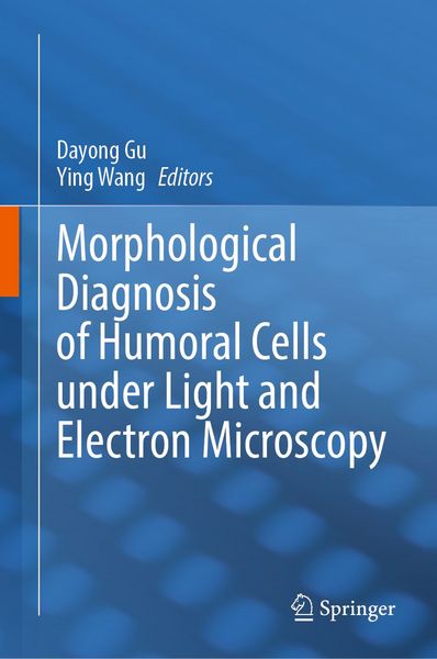
This book provides a detailed and practical guide to the morphological diagnosis of humoral cells using both light and electron microscopy. Electron microscopy compensates for the limited resolution of conventional examinations and plays a critical role in identifying tumors and resolving diagnostically challenging cases. Beginning with fundamental cell biology, the book analyzes cellular structures and ultrastructural features under electron microscopy and compares them with their counterparts under light microscopy. It further explains the relationships and key differences between the two imaging modalities through clinically relevant examples of abnormal cells. By linking light microscopic morphology to organelle level and structural characteristics revealed by electron microscopy, the book helps clinicians better understand the origins of observed cellular changes. Combining concise explanations with richly illustrated comparisons, this volume offers a comprehensive introduction to the morphology of body fluid cells. It serves as a valuable reference for laboratory physicians, laboratory technicians, and medical students.
Noch keine Bewertungen vorhanden
Verfassen Sie die erste Bewertung zu diesem Artikel
Helfen Sie anderen Kundinnen und Kunden durch Ihre Meinung.
Kurze Frage zu unserer Seite
Vielen Dank für Ihr Feedback
Wir nutzen Ihr Feedback, um unsere Produktseiten zu verbessern. Bitte haben Sie Verständnis, dass wir Ihnen keine Rückmeldung geben können. Falls Sie Kontakt mit uns aufnehmen möchten, können Sie sich aber gerne an unseren Kund*innenservice wenden.
zum Kundenservice