KI-Beratung Beta

Gutscheinbedingungen

**Gültig bis 11.11.2026 auf Spielzeug, Schreibwaren, Filme, Geschenke & Trends, Musik, tolino eReader & Zubehör, Hörbücher und Hörbuch-Downloads, nicht preisgebundene Bücher und Kalender online auf thalia.at und in der Thalia App. Einzelne Artikel können ausgeschlossen sein. Aufgrund der Buchpreisbindung sind deutschsprachige Bücher und eBooks ausgenommen. Zusätzlich ausgenommen sind Abos & Flatrates, Games, Geschenkkarten/-boxen, Shelfies, Software, Zeitschriften sowie einzelne Artikel von tonies®. Pro Einkauf einmal einlösbar. Click & Collect nur bei Onlinevorabzahlung möglich. Keine Barauszahlung. Nicht kombinierbar mit anderen Aktionen und Gutscheinen. Gutschein wird auf max. 500€ Bestellwert angerechnet. Nicht gültig für Versandkosten und Services.

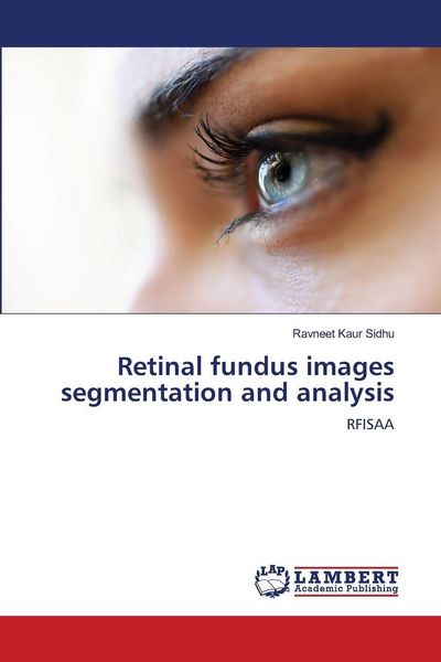
  • Produktbild: Retinal fundus images segmentation and analysis
  • Produktbild: Retinal fundus images segmentation and analysis
- 13%

Retinal fundus images segmentation and analysis RFISAA

13% sparen

38,99 € UVP 45,20 €

inkl. gesetzl. MwSt., Versandkostenfrei


Beschreibung

The cup to disk ratio (CDR) is the key indicator of this disease for detecting Glaucoma. It is observed that the CDR for healthy subjects lies in 0.233-0.306 range and for Glaucoma patients it lies in the range of 0.355-0.503. These values are in accord with the gold standards. Similarly for DR, in the...

Beschreibung

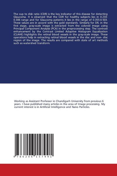
The cup to disk ratio (CDR) is the key indicator of this disease for detecting Glaucoma. It is observed that the CDR for healthy subjects lies in 0.233-0.306 range and for Glaucoma patients it lies in the range of 0.355-0.503. These values are in accord with the gold standards. Similarly for DR, in the first stage, gray-scale image is extracted from the colored image using Principal Component Analysis (PCA) in the preprocessing step. The contrast enhancement by the Contrast Limited Adaptive Histogram Equalization (CLAHE) highlights the retinal blood vessels in the gray-scale image. These operations help in extracting retinal blood vessels in the disc and non- disc region of the image. The results are compared with state of art methods such as watershed transform.

Produktdetails

Einband

Taschenbuch

Erscheinungsdatum

18.04.2023

Verlag LAP LAMBERT Academic Publishing
Seitenzahl

76

Produktdetails

Einband

Taschenbuch

Erscheinungsdatum

18.04.2023

Verlag LAP LAMBERT Academic Publishing
Seitenzahl

76

Maße (L/B/H)

22/15/0,6 cm

Gewicht

131 g

Sprache

Englisch

ISBN

978-620-6-15799-1

Herstelleradresse

VDM Verlag
Dudweiler Landstraße 99
66123 Saarbrücken
DE

Noch keine Bewertungen vorhanden

Verfassen Sie die erste Bewertung zu diesem Artikel

Helfen Sie anderen Kundinnen und Kunden durch Ihre Meinung.

Kundinnen und Kunden meinen

Bewertungen (0)

  • Produktbild: Retinal fundus images segmentation and analysis
  • Produktbild: Retinal fundus images segmentation and analysis