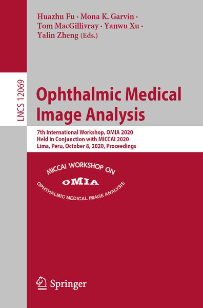
• Produktbild: Ophthalmic Medical Image Analysis
  • Produktbild: Ophthalmic Medical Image Analysis
Band 12069

Ophthalmic Medical Image Analysis 7th International Workshop, OMIA 2020, Held in Conjunction with MICCAI 2020, Lima, Peru, October 8, 2020, Proceedings

51,99 €

inkl. gesetzl. MwSt., Versandkostenfrei


Beschreibung

Produktdetails

Einband

Taschenbuch

Erscheinungsdatum

20.11.2020

Herausgeber

Huazhu Fu + weitere

Verlag

Springer

Seitenzahl

218

Maße (L/B/H)

23,5/15,5/1,3 cm

Gewicht

353 g

Auflage

1st ed. 2020

Sprache

Englisch

ISBN

978-3-030-63418-6

Beschreibung

Produktdetails

Einband

Taschenbuch

Erscheinungsdatum

20.11.2020

Herausgeber

Verlag

Springer

Seitenzahl

218

Maße (L/B/H)

23,5/15,5/1,3 cm

Gewicht

353 g

Auflage

1st ed. 2020

Sprache

Englisch

ISBN

978-3-030-63418-6

Herstelleradresse

Springer-Verlag KG
Sachsenplatz 4-6
1201 Wien
AT

Email: GPSR Kontakt

Noch keine Bewertungen vorhanden

Verfassen Sie die erste Bewertung zu diesem Artikel

Helfen Sie anderen Kundinnen und Kunden durch Ihre Meinung.

Kundinnen und Kunden meinen

Bewertungen (0)

  • Produktbild: Ophthalmic Medical Image Analysis
  • Produktbild: Ophthalmic Medical Image Analysis
  • Bio-Inspired Attentive Segmentation of Retinal OCT imaging.- DR detection using Optical Coherence Tomography Angiography (OCTA): a transfer learning approach with robustness analysis.- What is the optimal attribution method for explainable ophthalmic disease classification?.- DeSupGAN: Multi-scale Feature Averaging Generative Adversarial Network for Simultaneous De-blurring and Super-resolution of Retinal Fundus Images.- Encoder-Decoder Networks for Retinal Vessel Segmentation using Large Multi-Scale Patches.- Retinal Image Quality Assessment via Specific Structures Segmentation.- Cascaded Attention Guided Network for Retinal Vessel Segmentation.- Self-supervised Denoising via Diffeomorphic Template Estimation: Application to Optical Coherence Tomography.- Automated Detection of Diabetic Retinopathy From Smartphone Fundus Videos.- Optic Disc, Cup and Fovea Detection from Retinal Images using U-Net++ with EfficientNet Encoder.- Multi-level Light U-Net and Atrous Spatial Pyramid Poolingfor Optic Disc Segmentation on Fundus Image.- An Interactive Approach to Region of Interest Selection in Cytologic Analysis of Uveal Melanoma Based on Unsupervised Clustering.- Retinal OCT Denoising with Pseudo-Multimodal Fusion Network.- Deep-Learning-Based Estimation of 3D Optic-Nerve-Head Shape from 2D Color Fundus Photographs in Cases of Optic Disc Swelling.- Weakly supervised retinal detachment segmentation using deep feature propagation learning in SD-OCT images.- A framework for the discovery of retinal biomarkers in Optical Coherence Tomography Angiography (OCTA).- An Automated Aggressive Posterior Retinopathy of Prematurity Diagnosis System by Squeeze and Excitation Hierarchical Bilinear Pooling Network.- Weakly-Supervised Lesion-aware and Consistency Regularization for Retinitis Pigmentosa Detection from Ultra-widefield Images.- A Conditional Generative Adversarial Network-based Method for Eye Fundus Image Quality Enhancement.- Construction of quantitative indexes for cataract surgery evaluation based on deep learning.- Hybrid Deep Learning Gaussian Process for Diabetic Retinopathy Diagnosis and Uncertainty Quantification.