Beschreibung
Produktdetails
Einband
Gebundene Ausgabe
Erscheinungsdatum
15.04.2020
Abbildungen
<p>Approx. 3,000 illustrations (including full-color graphics)</p>, Illustrationen, nicht spezifiziert
Verlag
ElsevierSeitenzahl
928
Maße (L/B/H)
29,2/23,3/4,5 cm
Gewicht
2873 g
Sprache
Englisch
ISBN
978-0-323-66114-0
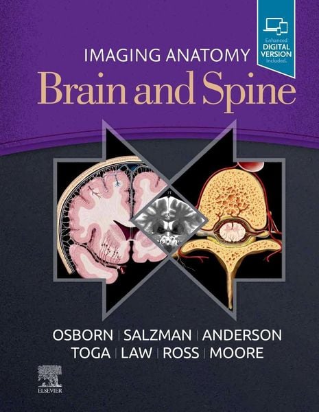
This richly illustrated and superbly organized text/atlas is an excellent point-of-care resource for practitioners at all levels of experience and training. Written by global leaders in the field, Imaging Anatomy: Brain and Spine provides a thorough understanding of the detailed normal anatomy that underlies contemporary imaging. This must-have reference employs a templated, highly formatted design; concise, bulleted text; and state-of- the-art images throughout that identify the clinical entities in each anatomic area.
- Features more than 2,500 high-resolution images throughout, including 7T MR, fMRI, diffusion tensor MRI, and multidetector row CT images in many planes, combined with over 300 correlative full-color anatomic drawings that show human anatomy in the projections that radiologists use
- Covers only the brain and spine, presenting multiplanar normal imaging anatomy in all pertinent modalities for an unsurpassed, comprehensive point-of-care clinical reference
- Incorporates recent, stunning advances in imaging such as 7T and functional MR imaging, surface and segmented anatomy, single-photon emission computed tomography (SPECT) scans, dopamine transporter (DAT) scans, and 3D quantitative volumetric scans
- Places 7T MR images alongside 3T MR images to highlight the benefits of using 7T MR imaging as it becomes more widely available in the future
- Presents essential text in an easy-to-digest, bulleted format, enabling imaging specialists to find quick answers to anatomy questions encountered in daily practice
- Includes the Expert Consult(TM) version of the book, allowing you to search all the text, figures, and references on a variety of devices
Noch keine Bewertungen vorhanden
Verfassen Sie die erste Bewertung zu diesem Artikel
Helfen Sie anderen Kundinnen und Kunden durch Ihre Meinung.
Kurze Frage zu unserer Seite
Vielen Dank für Ihr Feedback
Wir nutzen Ihr Feedback, um unsere Produktseiten zu verbessern. Bitte haben Sie Verständnis, dass wir Ihnen keine Rückmeldung geben können. Falls Sie Kontakt mit uns aufnehmen möchten, können Sie sich aber gerne an unseren Kund*innenservice wenden.
zum Kundenservice