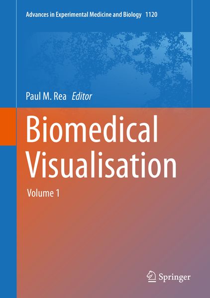
Biomedical Visualisation Volume 1
-
- Hardcover ausgewählt
- Taschenbuch
- eBook
-
Sprache:Englisch
-
Auflage:1st ed. 2019
- 1st edition 2020 149,99 €
- 1st ed. 2019 99,99 €
- 1st ed. 2019 99,99 €
- 1st ed. 2019 149,99 € ausgewählt
- 1st ed. 2019 149,99 €
- 1st ed. 2020 99,99 €
- 1st ed. 2020 149,99 €
- 1st ed. 2022 144,99 €
- 1st ed. 2021 129,99 €
- 1st ed. 2021 149,99 €
- 1st edition 2019 99,99 €
- 1st ed. 2023 144,99 €
- 1st ed. 2023 161,99 €
- 1st ed. 2023 175,99 €
149,99 €
inkl. gesetzl. MwSt.,
Beschreibung
Produktdetails
Einband
Gebundene Ausgabe
Erscheinungsdatum
09.04.2019
Abbildungen
XV, 65 illus., 42 illus. in color., schwarz-weiss Illustrationen, farbige Illustrationen
Herausgeber
Paul M. ReaVerlag
SpringerSeitenzahl
132
Maße (L/B/H)
26/18,3/1,4 cm
Gewicht
523 g
Auflage
1st ed. 2019
Sprache
Englisch
ISBN
978-3-030-06069-5
This edited volume explores the use of technology to enable us to visualise the life sciences in a more meaningful and engaging way. It will enable those interested in visualisation techniques to gain a better understanding of the applications that can be used in imaging and analysis, education, engagement and training.
The reader will be able to explore the utilisation of technologies from a number of fields to enable an engaging and meaningful visual representation of the life sciences. This use of technology-enhanced learning will be of benefit for the learner, trainer, in patient care and the wider field of education and engagement.
By examining a range of techniques in image capture (photogrammetery, stereophotogrammetry, microphotogrammetry and autostereoscopy), this book will showcase the wide range of tools we can use. Researchers in this field will be able to find something suitable to apply to their work to enhance user engagement through improved visual meansusing the technologies we have available to us today.It will highlight the uses of these technologies to examine many aspects of the human body, and enable improved ways to enhance visual and tactile learning, including 3D printing. By demonstrating co-design processes, working directly with the end-stage users (including patients), it will also highlight successes in adopting tools like hand motion tracking rehabilitation for patients with conditions like multiple sclerosis.
The book will also discuss the applications of immersive environments including virtual, augmented and mixed reality. The ultimate aim is to show how, by using these tools, we can enhance communication, mobile applications, health literacy and illustration of both normal and pathological processes in the body. By applying a wide range of tools and technologies, this volume will highlight the wide range of applications in education, training and learning both for students and faculty, butalso for patient care and education.
Therefore, the work presented here can be accessed by a wide range of users from faculty and students involved in the design and development of these processes, by examining the pedagogy around these technologies. Importantly, it presents material, which will be of benefit for the patient, engaging them to become more involved with techniques like physiotherapy.
Noch keine Bewertungen vorhanden
Verfassen Sie die erste Bewertung zu diesem Artikel
Helfen Sie anderen Kundinnen und Kunden durch Ihre Meinung.
Kurze Frage zu unserer Seite
Vielen Dank für Ihr Feedback
Wir nutzen Ihr Feedback, um unsere Produktseiten zu verbessern. Bitte haben Sie Verständnis, dass wir Ihnen keine Rückmeldung geben können. Falls Sie Kontakt mit uns aufnehmen möchten, können Sie sich aber gerne an unseren Kund*innenservice wenden.
zum Kundenservice