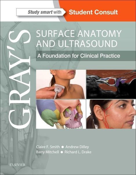
Gray's Surface Anatomy and Ultrasound A Foundation for Clinical Practice
-
- Taschenbuch ausgewählt
- eBook
-
Sprache:Englisch
48,99 €
inkl. gesetzl. MwSt.,
Beschreibung
Produktdetails
Einband
Taschenbuch
Erscheinungsdatum
25.08.2017
Abbildungen
317 figures (121 artwork, 108 ultrasound figures, 261 halftones), Illustrationen, nicht spezifiziert
Verlag
Elsevier Health ScienceSeitenzahl
216
Maße (L/B/H)
27,7/22,2/1 cm
Gewicht
620 g
Sprache
Englisch
ISBN
978-0-7020-7018-1
A concise, superbly illustrated (print + electronic) textbook that brings together a reliable, clear and up to date guide to surface anatomy and its underlying gross anatomy, combined with a practical application of ultrasound and other imaging modalities.
A thorough understanding of surface anatomy remains a critical part of clinical practice, but with improved imaging technology, portable ultrasound is also fast becoming integral to routine clinical examination and effective diagnosis.
This unique new text combines these two essential approaches to effectively understanding clinical anatomy and reflects latest approaches within modern medical curricula. It is tailored specifically to the needs of medical students and doctors in training and will also prove invaluable to the wide range of allied health students and professionals who need a clear understanding of visible and palpable anatomy combined with anatomy as seen on ultrasound.
- Concise text and high quality illustrations, photographs, CT, MRI and ultrasound scans provide a clear, integrated understanding of the anatomical basis for modern clinical practice
- Highly accessible and at a level appropriate for medical students and a wide range of allied health students and professionals
- Reflects current curriculum trend of heavily utilizing living anatomy and ultrasound to learn anatomy
- Supplementary video content showing live ultrasound scans and guided areas of surface anatomy to bring content to life and reflect current teaching and clinical settings
- An international advisory panel appointed to add expertise and ensure relevance to the variety of medical and allied health markets
- Inclusion of latest ultrasound image modalities
- Designed to complement and enhance the highly successful Gray's family of texts/atlases although also effective as a stand-alone or alongside other established anatomy resources
Noch keine Bewertungen vorhanden
Verfassen Sie die erste Bewertung zu diesem Artikel
Helfen Sie anderen Kundinnen und Kunden durch Ihre Meinung.
Kurze Frage zu unserer Seite
Vielen Dank für Ihr Feedback
Wir nutzen Ihr Feedback, um unsere Produktseiten zu verbessern. Bitte haben Sie Verständnis, dass wir Ihnen keine Rückmeldung geben können. Falls Sie Kontakt mit uns aufnehmen möchten, können Sie sich aber gerne an unseren Kund*innenservice wenden.
zum Kundenservice