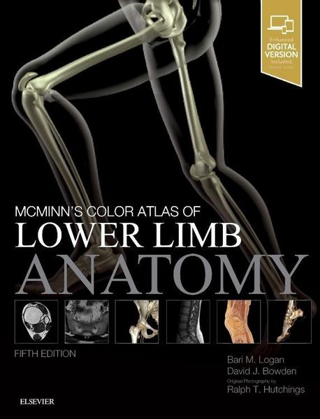
McMinn's Color Atlas of Lower Limb Anatomy
77,99 €
inkl. gesetzl. MwSt.,
Beschreibung
Produktdetails
Einband
Gebundene Ausgabe
Abbildungen
459 illustrations (265 in full color)
Verlag
Elsevier LTDMaße (L/B/H)
27,7/21,8/1,6 cm
Gewicht
800 g
Auflage
5. Auflage
Sprache
Englisch
ISBN
978-0-7020-7218-5
Understand foot, ankle and lower limb anatomy visually and clinically.
McMinn s Color Atlas of Lower Limb Anatomy is the ideal study aid or trusted visual reference for the range of students and practitioners who require a detailed understanding of the anatomy of the foot, ankle and lower limb. It provides you with the perfect grasp of all the important lower limb structures that are likely to be encountered in practice or in the anatomy lab. Superb images of dissections, osteology, radiographic and surface anatomy offer an unmatched view of muscles, nerves, skeletal structures, blood vessels and more. Figures are accompanied by concise notes and commentaries, as well as orientational artworks to help you locate the structure accurately on the body.
This updated fifth edition offers increased clinical relevance and features an entirely new chapter on Imaging of the Lower Limb, reflecting the very latest modalities and techniques. It also comes with the complete, enhanced eBook for the first time._
- All new state-of-the-art clinical imaging chapter - provides new insights and reflects anatomy as seen in modern practice
- Easily correlates essential anatomy with clinical practice - through over 200 high-quality photographs, combined with explanatory illustrations for more complex areas and concise, accompanying notes throughout
- Bonus information in Appendices - including practical guidance to administering nerve blocks accurately and effectively in the regional anaesthesia section
- Expert Consult(TM) eBook version included with purchase - this enhanced eBook experience allows you to search all of the text, figures, and references from the book on a variety of devices.
- Mostly life-size dissections and osteology - corresponds to what students/practitioners will see in the dissection lab or in real life
- Includes radiography and surface anatomy pictures - helps maximise clinical relevance (and necessary for modern courses)
- Orientational and explanatory artworks - helps the reader to position on the body
- Short accompanying text - expands on the illustrations and serves as study tool
- Numbered labels - helps facilitate self-testing
- Appendix containing key information on Skin, Muscles, Arteries and Nerves
Noch keine Bewertungen vorhanden
Verfassen Sie die erste Bewertung zu diesem Artikel
Helfen Sie anderen Kundinnen und Kunden durch Ihre Meinung.
Kurze Frage zu unserer Seite
Vielen Dank für Ihr Feedback
Wir nutzen Ihr Feedback, um unsere Produktseiten zu verbessern. Bitte haben Sie Verständnis, dass wir Ihnen keine Rückmeldung geben können. Falls Sie Kontakt mit uns aufnehmen möchten, können Sie sich aber gerne an unseren Kund*innenservice wenden.
zum Kundenservice