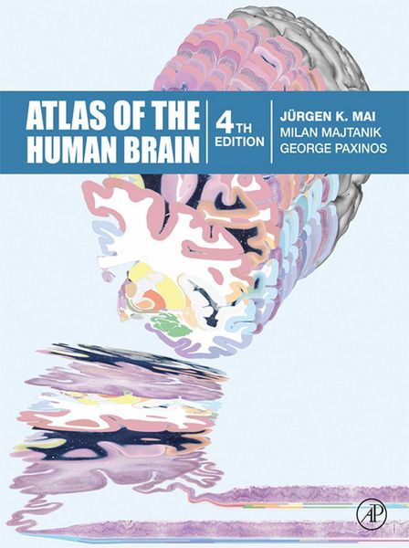
Atlas of the Human Brain
185,10 €
inkl. gesetzl. MwSt.Beschreibung
Produktdetails
Format
ePUB
Kopierschutz
Ja
Family Sharing
Nein
Text-to-Speech
Ja
Erscheinungsdatum
02.12.2015
Verlag
Elsevier Science & Techn.Seitenzahl
456 (Printausgabe)
Dateigröße
476893 KB
Auflage
4. Auflage
Sprache
Englisch
EAN
9780128028018
The fourth edition of Atlas of the Human Brain presents the anatomy of the brain at macroscopic and microscopic levels, featuring different aspects of brain morphology and topography. This greatly enlarged new edition provides the most detailed and accurate delineations of brain structure available. It includes features which assist in the new fields of neuroscience - functional imaging, resting state imaging and tractography. Atlas of the Human Brain is an essential guide to those working with human brain imaging or attempting to relate their observations on experimental animals to humans. Totally new in this edition is the inclusion of Nissl plates with delineation of cortical areas (Brodmann's areas), the first time that these areas have been presented in serial histological sections.
- Winner of the 2016 British Medical Association Award for Best Illustrated Text and previous edition winner of the Award of Excellence from the American Association of Publishers
- The contents of the Atlas of the brain in MNI stereotaxic space has been extensively expanded from 143 pages, showing 69 levels through the hemisphere, to 314 pages representing 99 levels
- In addition to the fiber-stained (myelin) plates, we now provide fifty new (Nissl) plates covering cytoarchitecture. These are interdigitated within the existing myelin plates of the stereotaxic atlas
- All photographic plates now represent the complete hemisphere
- All photographs of the cell- and fiber-stained sections have been transformed to fit the MNI-space
- Major fiber tracts are identified in the fiber-stained sections
- In the Nissl plates cortical delineations (Brodmann's areas) are provided for the first time
- The number of diagrams increased to 99. They were now generated from the 3D reconstruction of the hemisphere registered to the MNI- stereotaxic space. They can be used for immediate comparison between our atlas and experimental and clinical imaging results
- Parts of cortical areas are displayed at high magnification on the facing page of full page Nissl sections. Images selected highlight those areas which are thought to correspond with those published by von Economo and Koskinas (1925)
- A novel way of depicting cortical areal pattern is used: The cortical cytoarchitectonic ribbon is unfolded and presented linearly. This linear representation of the cortex enables the comparison of different interpretations of cortecal areas and allows mapping of activation sites
- Low magnification diagrams in the horizontal (axial) and sagittal planes are included, calculated from the 3D model of the atlas brain
Noch keine Bewertungen vorhanden
Verfassen Sie die erste Bewertung zu diesem Artikel
Helfen Sie anderen Kundinnen und Kunden durch Ihre Meinung.
Kurze Frage zu unserer Seite
Vielen Dank für Ihr Feedback
Wir nutzen Ihr Feedback, um unsere Produktseiten zu verbessern. Bitte haben Sie Verständnis, dass wir Ihnen keine Rückmeldung geben können. Falls Sie Kontakt mit uns aufnehmen möchten, können Sie sich aber gerne an unseren Kund*innenservice wenden.
zum Kundenservice