
Gutscheinbedingungen

**Gültig am 28.09.2026 auf fremdsprachige Bücher online auf thalia.at und in der Thalia App. Einzelne Artikel können ausgeschlossen sein. Ausgenommen sind preisgebundene Artikel & eBooks. Pro Einkauf einmal einlösbar. Click & Collect nur bei Onlinevorabzahlung möglich. Keine Barauszahlung. Nicht kombinierbar mit anderen Aktionen und Gutscheinen. Gutschein wird auf max. 500€ Bestellwert angerechnet. Nicht gültig für Geschenkkarten, Versandkosten und Services.

  • Produktbild: Information Processing in Medical Imaging
  • Produktbild: Information Processing in Medical Imaging
Band 1230

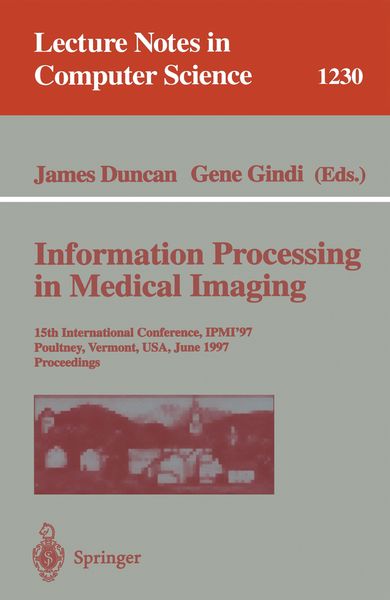
Information Processing in Medical Imaging 15th International Conference, IPMI'97, Poultney, Vermont, USA, June 9-13, 1997, Proceedings

51,99 €

inkl. gesetzl. MwSt., Versandkostenfrei


Beschreibung

Produktdetails

Einband

Taschenbuch

Erscheinungsdatum

21.05.1997

Abbildungen

XVI, 564 p.

Herausgeber

James Duncan + weitere

Verlag

Springer Berlin

Seitenzahl

564

Maße (L/B/H)

23,5/15,5/3,2 cm

Gewicht

867 g

Auflage

1997

Sprache

Englisch

ISBN

978-3-540-63046-3

Beschreibung

Produktdetails

Einband

Taschenbuch

Erscheinungsdatum

21.05.1997

Abbildungen

XVI, 564 p.

Herausgeber

Verlag

Springer Berlin

Seitenzahl

564

Maße (L/B/H)

23,5/15,5/3,2 cm

Gewicht

867 g

Auflage

1997

Sprache

Englisch

ISBN

978-3-540-63046-3

Herstelleradresse

Springer-Verlag KG
Sachsenplatz 4-6
1201 Wien
AT

Email: ProductSafety@springernature.com

Noch keine Bewertungen vorhanden

Verfassen Sie die erste Bewertung zu diesem Artikel

Helfen Sie anderen Kundinnen und Kunden durch Ihre Meinung.

Kundinnen und Kunden meinen

Bewertungen (0)

  • Produktbild: Information Processing in Medical Imaging
  • Produktbild: Information Processing in Medical Imaging
  • Automatic construction of eigenshape models by Genetic Algorithm.- Quadratic variation of deformations.- The softassign Procrustes matching algorithm.- Acoustic and elastic imaging to model disease-induced changes in soft tissue structure.- 2D intravascular E.I.T. using a non-iterative, non-linear reconstruction algorithm.- Optimal data types in optical tomography.- Using spatial-temporal images for analysis of gated cardiac tomographic data: The “M-Mode” revisited.- Automated segmentation of brain exterior in MR images driven by empirical procedures and anatomical knowledge.- Automated 3D segmentation using deformable models and fuzzy affinity.- Segmentation of medical image objects using deformable shape loci.- Mapping the cerebral sulci: Application to morphological analysis of the cortex and to non-rigid registration.- Bayesian detection of random signals on random backgrounds.- Approximate distributions for Maximum Likelihood and maximum a posteriori estimates under a Gaussian noise model.- Continuous Gaussian mixture modeling.- New statistical models for randoms-precorrected PET scans.- Registration of 3D medical images using simple morphological tools.- Deformation for image guided interventions using a three component tissue model.- Registration of abdominal CT and SPECT images using Compton scatter data.- Definition of volume transformations for volume interaction.- Nonlinear versus linear models in functional neuroimaging: Learning curves and generalization crossover.- Synthetic resampling methods for variance estimation in parametric images.- Space-time statistical model for functional MRI image sequences.- Jackknifing a normal database for regional cerebral blood flow SPECT obtained using principal components.- Generating 3-D cardiac material markersusing tagged MRI.- Identification of myocardial tags in tagged MR images without prior knowledge of myocardial contours.- An autofocus algorithm for the automatic correction of motion artifacts in MR images.- Measurement of global and regional cerebral volume changes by integrating boundary shifts between registered serial 3D MR scans.- Automatic detection of sulcal bottom lines in MR images of the human brain.- Segmentation and interpretation of MR brain images using an improved knowledge-based active shape model.- Gaussian random fields on sub-manifolds for characterizing brain surfaces.- Fuzzy logic approach to 3D magnetic resonance image segmentation.- Comparison of traditional brain segmentation tools with 3D self-organizing maps.- Finding the brain cortex using fuzzy segmentation, isosurfaces, and deformable surface models.- Statistical modelling of lines and structures in mammograms.- A thinning algorithm to extract medial lines from 3D medical images.- Setting the mind for intelligent interactive segmentation: Overview, requirements, and framework.- Segmentation of cerebral vessels and aneurysms from MR angiography data.- Computation of efficient patient specific models from 3-D medical images: Use in virtual endoscopy and surgery rehearsal.- In vivo analysis of trabecular bone architecture.- Automatic measurement of vertebral shape using Active Shape Models.- A hybrid approach for the detection of small airways disease from Computed Tomographic images.- Computer simulation of convection and diffusion effects on velocity estimations from X-ray contrast density time curves.- A comparison of retrospective intensity non-uniformity correction methods for MRI.- A robust and efficient algorithm for image registration.- Automatical adaption of the stereotactical coordinate system in brain MRI datasets.- Design of a statistical model of brain shape.- Automatic landmark identification using a new method of non-rigid correspondence.- Non-rigid curve correspondence for estimating heart motion.- Tracking of tagged MR images by Bayesian analysis of a network of quads.- Noninvasive skin imaging.- Modelling interreflections in a system based on the differential absorption of light (DAL) for the measurement of range images.- Quantitative estimation of scatterer spacing from backscattered ultrasound signals using the complex cepstrum.- Spatial compounding of 3-D ultrasound images.- Cone beam imaging of a section of a long object with a short detector.- New methods for SPECT imaging and pre-reconstruction restoration.- Reconstruction of the intracerebral vasculature from MRA and a pair of projection views.- Two alternative forced choice evaluation of vessel visibility increases due to zero-filled interpolation in MR angiography.- Bayesian detection with amplitude, scale, orientation and position uncertainty.